Rapid Change in the Pathology Lab – Choosing the Right Chemicals for Reducing Turnaround Time for Bone Marrow Biopsy
Medical technology, like all technology, changes at a rapid rate. What used to take a week or longer can now be done in just a day or two. And these changes include even the longest processes in the pathology lab.
Bone marrow trephine biopsy, an important tool in diagnosing and staging not just metastatic and primary bone tumors and hematologic malignancies, but also many systemic auto-immune and infectious diseases, has historically been one of those pathology processes with a lengthy turnaround time (TAT).
The prolonged processing time for bone marrow biopsy arises primarily because of the need to decalcify the mineralized or hard, bony structures. Decalcification may also be necessary in other tissues where dystrophic or metastatic calcification has occurred.
Fortunately for the patient and the entire medical team, new equipment and methods significantly reduce the TAT for bone marrow biopsy processing in the pathology lab.
The Development of Faster Alternatives
Newer alternatives to standard techniques in the pathology laboratory speed up decalcification without compromising the integrity of the sample.
The first of these has actually been in use for decades… the microwave.r alternatives to standard techniques in the pathology laboratory speed up decalcification without compromising the integrity of the sample.
There’s evidence that subjecting the tissue to microwaves speeds decalcification time by hours… making it anywhere from 20 to 26 percent faster. Interestingly, the microwave effect works regardless of the solution’s temperature. In a study by Tinling et al, decalcification time was reduced even when the temperature of the solution was maintained at 20°C.[1]
It goes without saying, but we’ll say it anyway… only laboratory-grade microwaves should ever be used in the lab. Along with a whole lot of other bells and whistles (such as stirring the reagent), lab-grade microwaves are made to vent toxic fumes.
Another technique that can decrease the decalcification time is carefully increasing the temperature while circulating or agitating the solution around the samples. Decalcification at 45°C has been shown to significantly reduce the time required to demineralize calcified tissue.[2] The agitation or stirring keeps the bone sample in contact with fresh decalcifier constantly, helping to speed up decalcification.
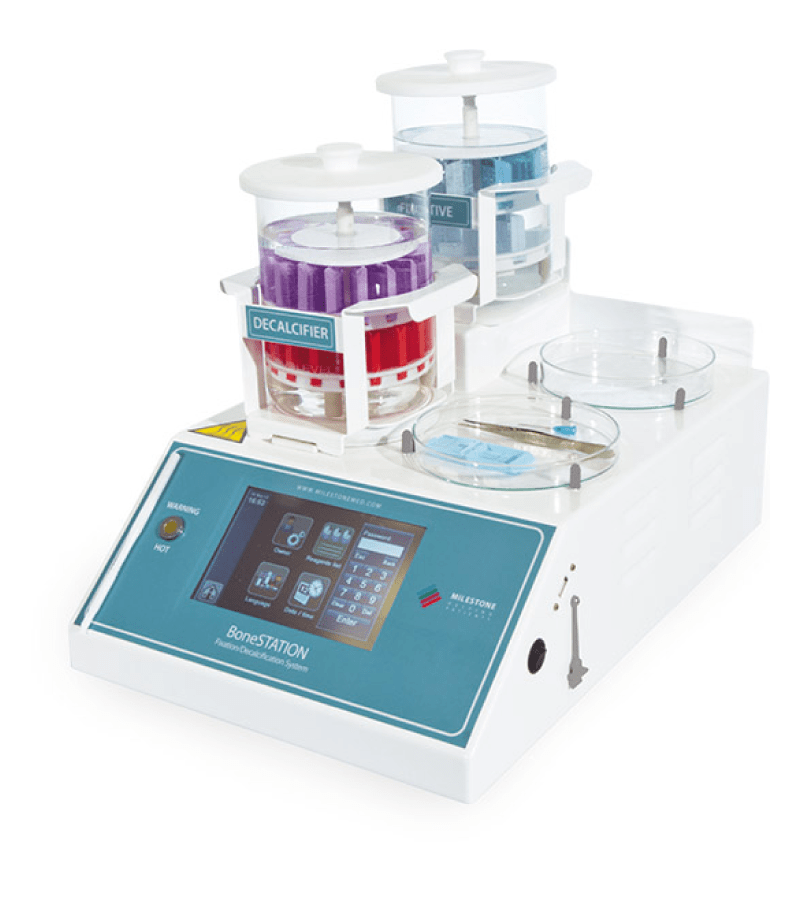
 Among the latest technology is the BoneSTATION by Milestone Medical. The BoneSTATION uses infrared to apply controlled heat while stirring the decalcifier around the tissue cassettes. It has a separate platform, also with magnetic stirring, for the fixation step. The BoneSTATION will tolerate even strong mineral acids for those who choose to use them, and its cover prevents fumes from escaping into the air.
Depending on which equipment is being used (microwave, BoneSTATION, or perhaps something else), up to 45 cassettes can be processed in one batch. Decalcification can be completed in around two hours.
Once a decision is made on which equipment to use, the chemicals need to be chosen… namely the fixative and decalcifier.
Fixative Options for Bone Marrow Trephine Biopsy
In order to preserve the samples for immunohistochemistry, formalin-based fixatives are preferred by most pathology labs.
The most common fixatives for bone marrow trephine biopsy are 10% neutral buffered formalin and acetic acid-zinc-formalin (AZF). According to the Hammersmith Protocol, formalin alone is not as good at preserving morphology and antigens for immunohistochemistry as zinc-containing fixatives. For this reason, they prefer acetic acid-zinc-formalin (AZF), with both zinc and formalin, for bone marrow trephine biopsy.[3]
Both AZF and formalin alone take about the same amount of time… approximately six hours depending on the sample size. Common practice is to allow samples to remain in the fixative overnight.
Faster fixatives are available, but they can damage morphology, nucleic acids, and antigens. Read more about fixatives here.
Choosing the Right Decalcifier for Improving Turnaround Time
We talked earlier about the need to decalcify bone marrow trephine biopsy samples. Without this step, it’s difficult to get good quality sections because of the hard, mineralized bones.
Inorganic strong acids will work quickly to decalcify, however, while they work for the decalcification of bone, the use of nitric or hydrochloric acid as a fast decalcifier isn’t the best option for preserving tissue morphology. The acids damage DNA and RNA as well as not being a viable option for some protein assays.[4]
That leaves two good options—EDTA and formic acid.
EDTA is recommended by the International Council for Standardization in Hematology (ICSH). EDTA preserves antigens, DNA, RNA, and proteins. It allows for molecular techniques including NGS and FISH assays to be carried out successfully.[5]
The Hammersmith Protocol calls for a solution of 10% formic acid and 5% formaldehyde.
Standard timing without the aid of the technology we mentioned here is anywhere from six hours and up. That can be brought down to just two hours depending on tissue size and the methods and equipment used.
Whichever your lab chooses, reduced TAT is a benefit to all.
Check out our selection of fixatives at PathSUPPLY.com or reach out to your PathSUPPLY rep for your pathology lab supply needs.
PathSUPPLY – helping those who help patients. Call us at 1-800-631-3556.
[1] TINLING, S. P., GIBERSON, R. T. and KULLAR, R. S. (2004), Microwave exposure increases bone demineralization rate independent of temperature. Journal of Microscopy, 215: 230-235. doi:10.1111/j.0022-2720.2004.01382.x
[2] Kapila SN, Natarajan S, Boaz K, Pandya JA, Yinti SR. Driving the Mineral out Faster: Simple Modifications of the Decalcification Technique. J Clin Diagn Res. 2015;9(9):ZC93–ZC97. doi:10.7860/JCDR/2015/14641.6569
[3] Naresh KN, Lampert I, Hasserjian R, et al. Optimal processing of bone marrow trephine biopsy: the Hammersmith Protocol. J Clin Pathol. 2006;59(9):903–911. doi:10.1136/jcp.2004.020610
[4] Veena M. Singh, Ranelle C. Salunga, Vivian J. Huang, Yen Tran, Mark Erlander, Pam Plumlee, Michael R. Peterson,
Analysis of the effect of various decalcification agents on the quantity and quality of nucleic acid (DNA and RNA) recovered from bone biopsies, Annals of Diagnostic Pathology, Volume 17, Issue 4, 2013, Pages 322-326, ISSN 1092-9134, https://doi.org/10.1016/j.anndiagpath.2013.02.001
[5] Peter Ntiamoah MPH, George H Ayob MBBCH, Richard R Clarke, Khedoudja Nafa PhD, Paulo A Salazar BS, Agnes Viale PhD, Ahmet Dogan MD PhD, Meera Hameed MD, Memorial Sloan Kettering Cancer Center, EDTA-Based Decalcification of Bone and Bone Marrow – Ideal Tool for Protein and Nucleic Acid Preservation – A Pilot Study
significantly reduce the time required to demineralize calcified tissue.[1] The agitation or stirring keeps the bone sample in contact with fresh decalcifier constantly, helping to speed up decalcification.
